
How to Treat Calcium Blocks in the Heart with Newer Techniques
If you or a loved one have been told that an angioplasty cannot be completed due to "hardened" or "calcified" blockages, it does not mean you are out of options.
7/19/20263 min read
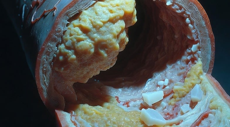
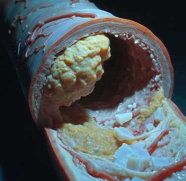
Severe calcium deposits in the coronary arteries can act like armor, preventing standard angioplasty balloons and stents from expanding properly to restore blood flow. Modern interventional cardiology utilizes advanced plaque-modification technologies—such as Intravascular Lithotripsy (IVL) and high-speed atherectomy—to safely crack or sand down this calcified buildup. These newer, minimally invasive techniques achieve excellent stent expansion and restore optimal circulation with far fewer complications than traditional, high-pressure methods.
What are "calcium blocks" in the heart, and why are they dangerous?
When plaque builds up inside the coronary arteries over decades, it undergoes a chemical transformation. Over time, calcium minerals deposit into these fatty blockages, turning soft plaque into a stone-like substance.
Think of normal arterial blockages like a buildup of soft butter inside a pipe; a standard angioplasty balloon can easily push it aside. Calcified blockages, however, are like solid concrete.
If an interventional cardiologist encounters severe calcium during a procedure, a standard balloon may burst without making a dent. Forcing a stent into an unprepared, calcified artery leads to "stent underexpansion". If the stent cannot open fully, the risk of the artery closing up again (restenosis) or forming sudden blood clots increases significantly.
What is Intravascular Lithotripsy (IVL), and how does it crack heart calcium?
One of the most revolutionary advancements in structural and interventional cardiology is Intravascular Lithotripsy (IVL). Borrowed from urology—where sonic shockwaves have been used for decades to break up kidney stones—IVL delivers localized acoustic pressure waves directly into the heart's arteries.
The Technology: A specialized balloon catheter is guided directly to the calcified blockage using a standard guidewire.
The Mechanism: Once in place, the balloon is inflated to a very low pressure just to make contact with the artery wall. Miniature emitters inside the balloon then release pulsatile sonic waves.
The Result: These acoustic waves travel harmlessly through the soft, elastic tissue of the blood vessel but create multiple microfractures when they hit the brittle calcium.
Because IVL fractures both superficial (surface) and deep medial calcium within the vessel wall, it completely changes the compliance of the artery. The rigid "concrete" is turned into cracked gravel, allowing a drug-eluting stent to expand flawlessly and safely.
How do Rotational and Orbital Atherectomy work for severe calcification?
For specific types of complex, rock-hard blockages—especially those so tight that an IVL balloon cannot even cross them—automated advanced drilling or sanding techniques are used.
Rotational Atherectomy (Rotablation)
This technique uses a tiny, diamond-encrusted elliptical burr that spins at up to 140,000+ RPM. It acts like a high-speed clinical drill, cutting through the hard calcium while shaving it down into microscopic particles smaller than red blood cells, which safely wash away in the bloodstream. It is particularly effective for opening up exceptionally narrow, uncrossable blockages.
Orbital Atherectomy
A newer evolution of the drilling concept, orbital atherectomy features a diamond-coated crown mounted eccentrically on a spinning shaft. Instead of drilling straight ahead, it travels in a controlled orbital path inside the artery, creating a widening circle of ablation.
Bidirectional Sanding: It can shave calcium moving both forward and backward, drastically reducing the risk of the device getting stuck in a tight blockage.
Tissue Protection: Using the laws of centrifugal force, it selectively sands away rigid, non-compliant calcium while deflecting safely away from healthy, elastic arterial tissue.
How do cardiologists choose the right technique?
Not all heart calcium is created equal, and modern cardiac care relies heavily on intravascular imaging tools like Intravascular Ultrasound (IVUS) or Optical Coherence Tomography (OCT) to look inside the vessel wall before deciding on a treatment strategy.
Feature / ScenarioIntravascular Lithotripsy (IVL)Orbital / Rotational AtherectomyPrimary MechanismSonic shockwaves that fracture deep & superficial calcium.High-speed diamond crowns that sand down surface calcium.Best Used ForCircumferential (360-degree) deep calcium, large vessels, and bifurcations.Tightly occluded, pinpoint blockages where a balloon cannot pass.Vessel Safety ProfileExtremely low risk of perforation; leaves soft tissue completely untouched.Requires precise wire bias control to prevent spinning burr complications.Procedural EaseBalloon-based platform with a short learning curve.Specialized advanced equipment requiring extensive technical training.
In many complex cases, a hybrid approach is utilized. A brief round of atherectomy is performed first to create a small channel through an uncrossable calcium block, followed by IVL to fracture the deep walls of the rock, ensuring a perfect landing zone for the final stent.
When should you consult a specialist about calcified heart blockages?
If you or a loved one have been told that an angioplasty cannot be completed due to "hardened" or "calcified" blockages, it does not mean you are out of options. Traditional balloon procedures are no longer the limit of care. Advanced structural heart teams possess the specialized tools necessary to safely shatter the toughest calcium armor and completely restore health to the heart.
If you would like an expert second opinion regarding complex coronary interventions, calcified lesion management, or custom preventative cardiology care, please reach out through our dedicated patient consultation channels:
In-Person Appointments (Meitra Hospital, Kozhikode): Connect directly with the Centre for Heart & Vascular Care.
Private Practice (SA Heart and Vascular Clinic, Thondayad Junction, Kozhikode): For localized clinical consultations.
International Expat Portal & Online Second Opinions: Schedule a dedicated digital video consultation from anywhere globally.
Location
SA clinic:
2 nd floor , Pentagon healthcare Thondayad, Kozhikode, Kerala 673017
MEITRA HOSPITAL:
Karaparamba, Mini Bypass Rd, Kunduparampa, Edakkad, Kozhikode, Kerala 673005
Hours
SA clinic 9:00-11:00
Meitra Hospital 11:30-5:00
Booking
SA Clinic: 00918138001022
MEITRA hopsital: 04957123456